paper · preprint, published here in full
Two independent measured datasets support the direction of an iPSC-cardiomyocyte repolarization-risk model
Daniel Reda
Perturb Bio, Inc.
Against Blinova et al. 2018, 112 matched drug-dose points give a Spearman rank correlation of 0.67 and direction agreement on 89% of measurable points (82 of 92). Against Lee et al. 2025, direction agrees on 9 of 10 anchored drugs. This is the preprint text in full.
abstract
An in-silico repolarization-risk model used in a commercial mechanism read was validated in two ways against two independent published human iPSC-cardiomyocyte datasets. Against the Blinova et al. (2018) multi-lab beat-rate study, the model's predicted repolarization direction matched measurement on 82 of 92 measurable drug-dose points (89%), with a Spearman rank correlation of 0.67 across 112 matched points; excluding collapse points the correlation was 0.62. The model collapsed on six drugs, and Blinova et al. annotated EAD for each of them. Against the Lee et al. (2025) 28-drug FPDc screen, the model's direction agreed on 9 of 10 anchored drugs. The two agreements establish repolarization direction and, against Blinova, rank order, and they bound the validation's scope: waveform and magnitude equivalence, and the mechanism ranking, sit outside it.
1 · The question
The model under test is a human iPSC-cardiomyocyte action-potential model (Kernik et al. 2019) run forward from a panel of ion-channel block to a repolarization-risk score, and run in reverse from a measured repolarization phenotype to a ranked set of conductance mechanisms. The forward direction has been scored against the CiPA reference classes on 28 drugs and the result published. The question here is narrower: does the model's direction of repolarization change, and its ordering across drugs and doses, agree with what two independent labs measured?
Two datasets give the answer. Blinova et al. (2018) provide matched drug-dose points from a multi-site study on a single iPSC-CM line. Lee et al. (2025) provide a 28-drug screen, reported as a figure heatmap, from which ten drugs could be anchored with confidence.
2 · Against Blinova et al. 2018
We matched every drug-dose point the two studies share and compared the sign of the model's APD90 change with the sign of the measured FPD change. Of 92 measurable points, the model agreed on 82 (89%). Removing points where the model collapsed gave a correlation of 0.62 across the remainder. Including all matched points, the Spearman rank correlation was 0.67.
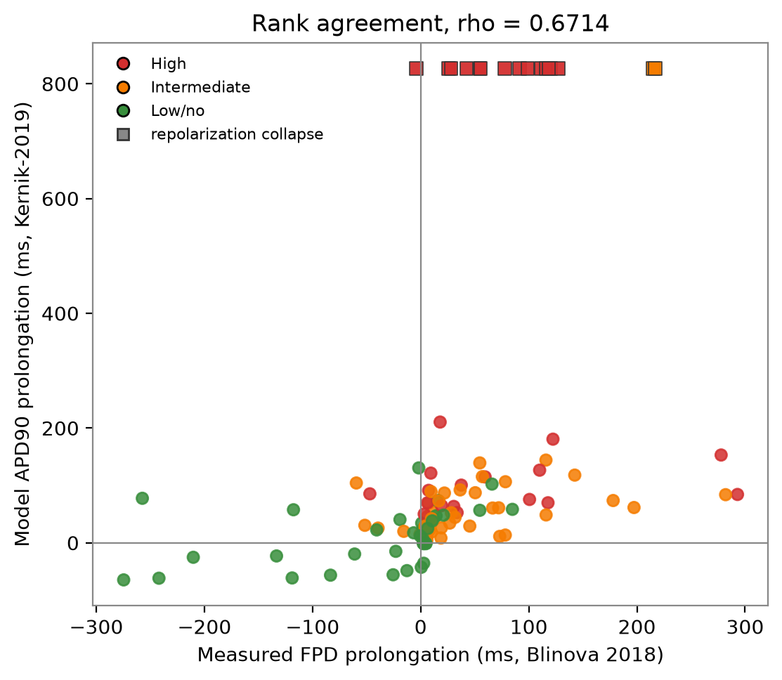
Figure 1. Model APD90 prolongation against measured FPD prolongation for 112 matched drug-dose points from Blinova et al. 2018. Squares mark points where the model collapsed; Blinova annotated EAD at each.
The model collapsed, failing to repolarize within the simulation window, on six drugs. Blinova et al. annotated EAD for all six. There were no false collapses. As a detector of EAD-bearing drugs among the panel's 27, the model's collapse caught six, a sensitivity of 22%, and its precision was 100%.
The model over-prolongs in absolute milliseconds. Its APD90 values run high against the measured FPD values. The agreement is in direction and in rank; magnitude is outside this comparison's scope.
3 · Against Lee et al. 2025
Lee et al. report a 28-drug iPSC-CM FPDc screen as a heatmap, so the published figure yields a partial rank read. We anchored ten drugs we could read with confidence and compared the direction of the model's response to the direction the screen shows.
direction agreement, ten anchored drugs
| drug | Lee et al. (measured) | model | agree |
|---|
| quinidine | prolong | prolong | yes |
| dofetilide | prolong | prolong | yes |
| droperidol | prolong | prolong | yes |
| ibutilide | prolong | prolong | yes |
| sotalol | prolong | prolong | yes |
| ranolazine | no prolong | no prolong | yes |
| nifedipine | shorten | shorten | yes |
| nitrendipine | shorten | shorten | yes |
| diltiazem | shorten | shorten | yes |
| verapamil | shorten | prolong | no |
Nine of ten drugs agree. The counterintuitive case is ranolazine: two measurements and the model agree it does not prolong, despite its block of hERG. The model's own hERG block would suggest prolongation, and the agreement says otherwise. The model carries no distinct late-sodium current, so the run has no late-sodium route. It reads the correct direction anyway, and no mechanism explains the reading.
The single disagreement is verapamil. Lee et al. measure a shortening; the model predicts a prolongation. Verapamil blocks calcium and hERG together, and this is the third independent place the model's calcium-block limitation has been confirmed. It is stated in the read.
4 · What is established
- Repolarization direction agrees with two independent datasets. 82 of 92 measurable points against Blinova; 9 of 10 anchored drugs against Lee.
- Rank order agrees with Blinova. Spearman rank correlation 0.67 across 112 matched points.
- The calcium-block limitation is confirmed a third time. Verapamil is the sole direction miss, and the cause is known.
Measured failure modes, stated:
- The population-confidence signal is overconfident. Its top band reports a mean confidence of 0.99 against an observed accuracy of 0.63. It carries ordering only, and the read treats it as a ranking, with the observed accuracy published alongside.
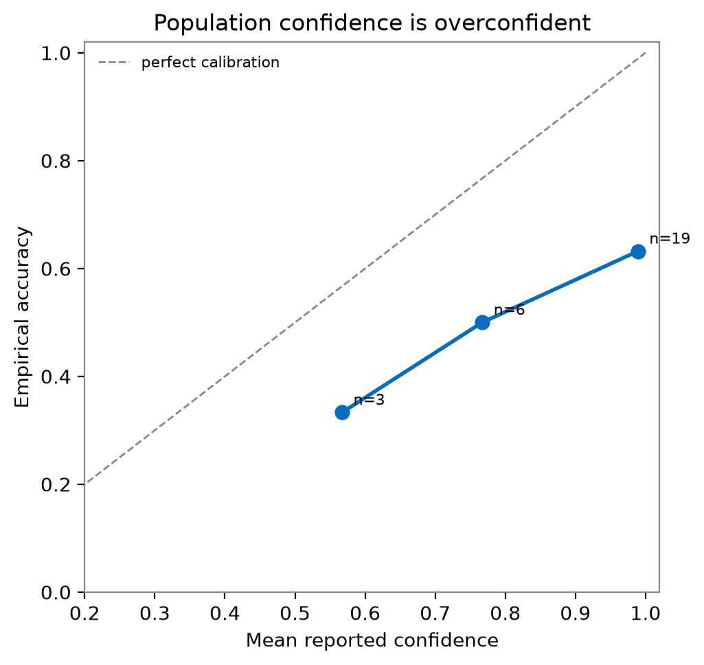
Figure 2. Reliability of the population-confidence signal. Points fall below the identity line, showing the reported confidence runs ahead of observed accuracy.
- Adding IKs did nothing. The 28-drug accuracy is unchanged at 17 of 28, and no drug changes class. A null result, published.
- Adding a late-sodium current lowered validation accuracy. It is a negative result, and we publish it.
5 · Limits
This validation scores sign and rank, so its scope is direction and order. Shape sits outside it; the model over-prolongs in absolute milliseconds, so magnitude sits outside it too. The Lee figure yields a partial rank read, so a full 28-drug order against Lee stays outside. The mechanism ranking, the product's use, stays outside as well; testing it needs matched wet mechanism data. A fast mechanism answer is a bench decision aid. A regulatory submission is a separate, much longer path.
validation scope: direction and rank
- The mechanism ranking is a separate claim. This paper tests direction and rank, the inputs to the read. Testing the ranked mechanisms against wet-lab mechanism needs matched wet data, and this study ran with the direction and rank datasets.
- Funding and interest. This work received no funding. The author is the sole founder of Perturb Bio, which sells the in-silico mechanism read the model underlies.